Figures & data
Figure 1. Hypoxia induces apoptosis and autophagy of PC-12 cells. (A) Cell viability was studied by MTT assay. (B) Flow cytometry analysis was performed for cell apoptosis detection. (C,D) Relative expression of apoptotic proteins was detected by Western blot. (E,F) The accumulated levels of p62 and LC3-II/LC3-I were detected by Western blot. Data are shown as mean ± SD. **p < .01; ***p < .001.
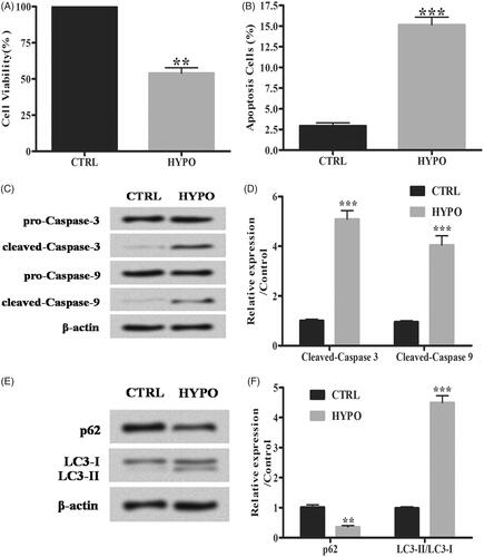
Figure 2. 6-Gingerols (6G) alleviates hypoxia induced apoptosis and autophagy in PC-12 cells. (A,B) Cell viability was detected by MTT assay. (C) Flow cytometry analysis was performed for cell apoptosis detection. (D,E) Relative expression of apoptotic factors was detected by Western blot. (F,G) The accumulated levels of p62 and LC3-II/LC3-I were detected by Western blot. Data are shown as mean ± SD. *p < .05; **p < .01; ***p < .001.
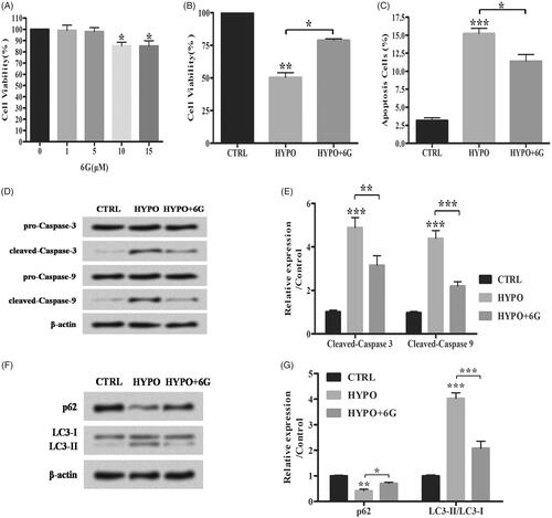
Figure 3. 6-Gingerols (6G) increases miR-103 expression in PC-12 cells. (A) Relative expression of miR-103 was detected by qRT-PCR. Data are reported as mean ± SD. *p < .05.
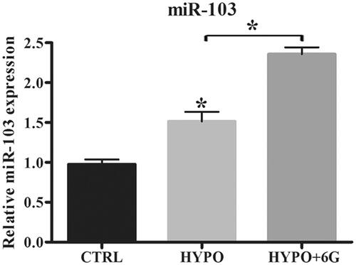
Figure 4. 6-Gingerols (6G) alleviates hypoxia induced injury by up-regulating miR-103 in PC-12 cells. (A) Relative expression of miR-103 was detected by qRT-PCR. (B) Cell viability was examined by MTT assay. (C) Flow cytometry analysis was performed for cell apoptosis detection. (D,E) Relative expression of apoptosis-related proteins was detected by Western blot. (F,G) Relative expression of p62 and LC3/LC3-I ratio were detected by Western blot. Data are reported as mean ± SD. *p < .05; **p < .01; ***p < .001.
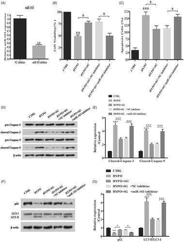
Figure 5. Overexpression of Bcl2/BNIP3 induces apoptosis and autophagy of PC-12 cells. (A,B) Relative expression of BNIP3 was detected by Western blot. (C) Results from dual luciferase activity assay. Data are reported as means ± SD. *p < .05.
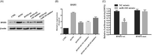
Figure 6. 6-Gingerols (6G) inhibits p38 MAPK and JNK signalling pathways through down-regulation of BNIP3. (A,B) Relative expression of BNIP3 was detected by Western blot. (C) Flow cytometry analysis was performed for cell apoptosis detection. (D,E) Relative expression of apoptosis-related factors was detected by Western blot. (F,G) Relative expression of p62 and LC3/LC3-I ratio were detected by Western blot. Data are reported as mean ± SD. *p < .05; **p < .01; ***p < .001.
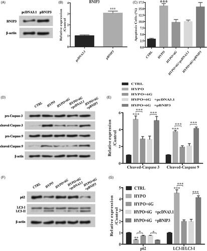
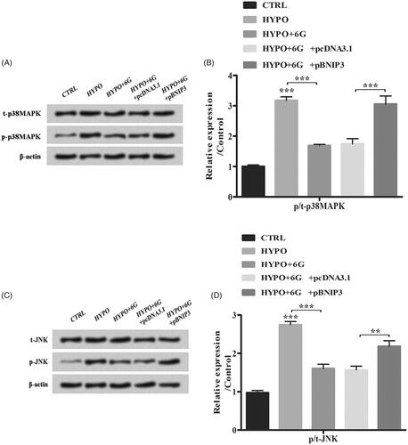
Figure 7. 6-Gingerols (6G) inhibits p38 MAPK and JNK signalling pathways through down-regulation of BNIP3. (A,B) Relative expression of t-p38MAPK and p-p38MAPK was measured by Western blot. (C,D) Relative expression of t-JNK and p-JNK was detected by Western blot. Data are reported as mean ± SD. **p < .01; ***p < .001.