Figures & data
Figure 1. Elevated level of CGRP was observed in HO following spinal cord injury. (A) The X ray imaging demonstrated the formation of callus 2 and 4 weeks after modelling. (B,C) The H&E and Alcian Blue staining showed the formation of cartilage and bone-like tissues. (D) The chondrogenic and osteogenic genes also elevated significantly at day 14. (E) An increased level of CGRP was observed in muscle samples at 14 days after modelling.
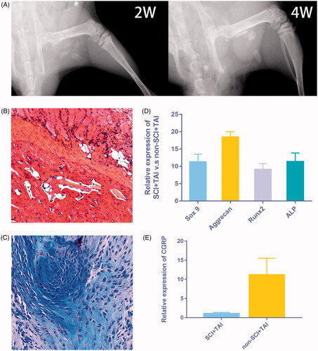
Figure 2. Directly injection of CGRP can promote callus formation in vivo. (A) The cross-sectional area of the callus is much larger in the SCI + TAI group than in the control (p = .003). (B) Directly injection of CGRP was able to induce the formation of cartilage/bone like tissues as shown by X ray of 2w post injection. (Red arrow: no cartilage/bone like tissue forms after PBS injection; Blue arrow: cartilage/bone like tissues forms after CGRP injection.) (C) The Alcian Blue staining of the samples 2w post injection. (Green arrow: limited areas of inflammatory infiltration; Yellow arrow: Chondrocytes formation after CGRP injection.)
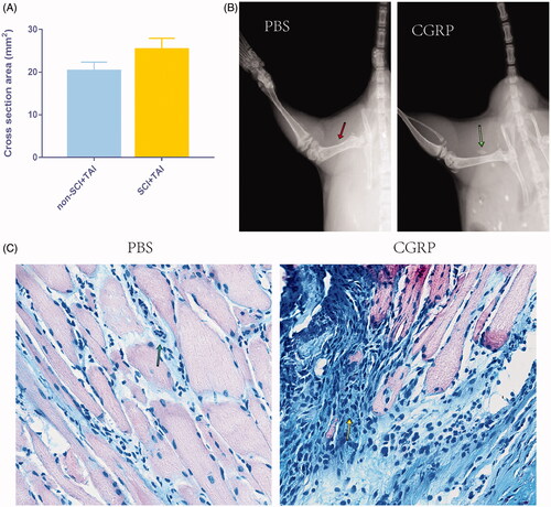
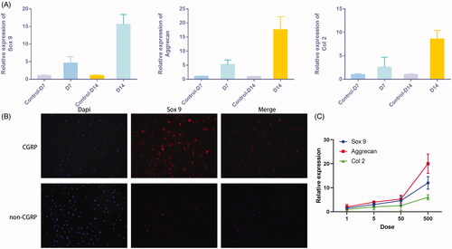
Figure 3. CGRP stimulates chondrogenic differentiation of FAPs in vitro. (A) Additional CGRP was able to induce chondrogenic differentiation of FAPs, as shown by increased expression of Sox9, Aggrecan, and Col II. (B) Immunofluorescence of Sox 9 was more significant with additional CGRP. (C) The expression of chondrogenic genes was in a dose-dependent manner with the dose of the additional CGRP.