Figures & data
Figure 1. Target muscles before and after intramuscular injections of tracers. (A) Before injection. (B) After injection: target muscles were completely Coloured.
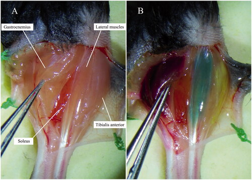
Figure 2. Harvest of the spinal cord (spinal nerve roots remained).
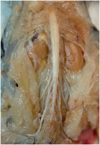
Table 1. Properties of the retrograde tracers and setups of lasers and filters of Zeiss LSM 780 confocal microscope.
Figure 3. Spinal cord before and after optical clearing procedure. (A) Before clearing. (B) After clearing.
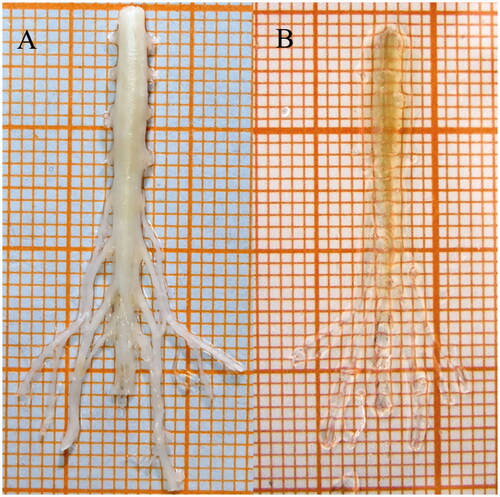
Figure 4. MIP images of single track. (A) Tibialis anterior motor neurons labelled by FG. (B) Soleus motor neurons labelled by CTb-488. (C) Gastrocnemius motor neurons labelled by CTb-488. (D) Lateral muscle motor neurons labelled by CTb-647.
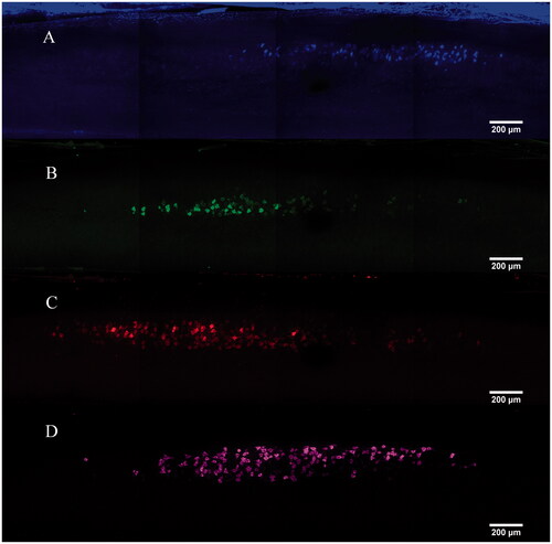
Figure 5. MIP images of multi-tracks in a holistic view [motor neurons innervating tibialis anterior (blue), gastrocnemius (red), soleus (green), lateral muscle (magenta)]. White arrows: dendrites or axons around the soma.
![Figure 5. MIP images of multi-tracks in a holistic view [motor neurons innervating tibialis anterior (blue), gastrocnemius (red), soleus (green), lateral muscle (magenta)]. White arrows: dendrites or axons around the soma.](/cms/asset/15a36e65-0753-431e-b498-17917771890f/ianb_a_1687493_f0005_c.jpg)
Table 2. Imaging results of motor neuron pools labelled by the four tracers.
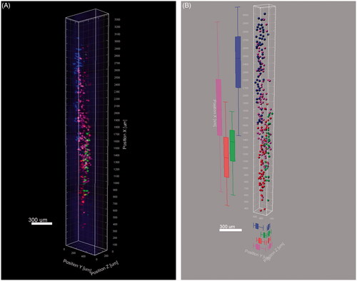
Figure 6. Three dimensional reconstruction of the labelled neurons. (A) Snapshot of the 3D reconstruction. (B) Vantage analysis of the four motor neuron pools.