Figures & data
Figure 1 Images (H&E) of the lung showing 4 μm thick (A) and 300 μm thick (B–F) sections at ×40 magnification (A and B) and ×100 magnification (C–F).
Notes: The polyhedral complex of alveoli is composed of a framework (C, arrows) and an alveolar wall. We designated the face of the alveolar polyhedron as the FU (D–F, white dashed lines). We categorized FUs into four types based on their morphological characteristics: 1) FUs without an alveolar wall (white dashed lines with an asterisk in D), 2) FUs with fragments of an alveolar wall (white dashed lines in E), 3) FUs with an intact alveolar wall with pores of Kohn (white dashed lines with a single dagger in D and white dashed lines in F), and 4) FUs with an intact alveolar wall without pores of Kohn (white dashed lines with a double dagger in D). The framework is thicker than the cut edges of the alveolar wall (E, arrowheads). The area of each pore and the sum of the area of all pores seen in one FU were evaluated by the percentage area of the FU (F, solid black line tracing and arrows). The black and white scale bars indicate 200 μm.
Abbreviations: FU, framework unit; H&E, hematoxylin and eosin.
Abbreviations: FU, framework unit; H&E, hematoxylin and eosin.
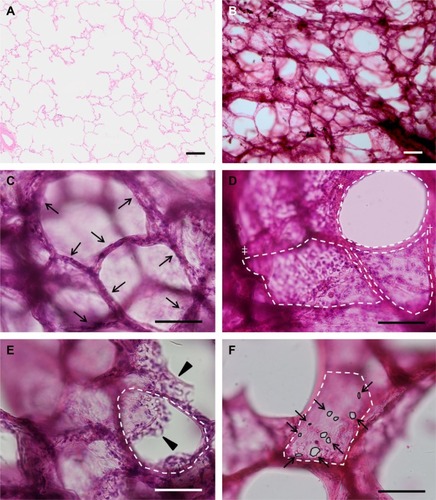
Table 1 Characteristics of the cases
Table 2 Comparison of FUs and pores of Kohn between groups with and without emphysema
Table 3 Bivariable and multivariable regression analysis on DLCO
Table 4 Bivariable and multivariable regression analysis on DLCO/VA
Table 5 Bivariable and multivariable regression analysis on RV
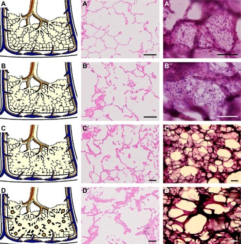
Figure 2 Schemas (A–D) and images of an ordinary H&E slide (A′–D′) and an H&E thick section (A″–D″) on possible progression of pulmonary emphysema.
Notes: Images A′ and B′ are at ×50 magnification. Images A″ are B″ are at ×80 magnification. Images C′, C″, D′, and D″ are at ×30 magnification. According to our results, the morphological progression of pulmonary emphysema is proposed as follows: alveoli with a few tiny pores of Kohn in the normal state (A–A″) starts to show an increase in the number and size of pores of Kohn, resulting in vulnerability of the alveoli (B–B″); the framework structure and alveoli merge and break down because of mechanical stress and/or inflammation, inducing traction of lung tissue (C–C″); and remodeling of acini resulting in enlarged air spaces of emphysema (D–D″). The pathological slide of images A′–D′ and the thick sections of image A″–D″ were processed from the same tissue block of an identical case. The black and white bars indicate 200 μm.
Abbreviation: H&E, hematoxylin and eosin.
Abbreviation: H&E, hematoxylin and eosin.