Figures & data
Figure 1 TUNEL staining for apoptotic cells in retinas at 2 weeks after intravitreal injection.
Notes: (A) Low group: intravitreal injection of 2.5 mg/0.1 mL PLGA/PLA microspheres; (B) medium group: intravitreal injection of 5 mg/0.1 mL PLGA/PLA microspheres; (C) high group: intravitreal injection of 10 mg/0.1 mL PLGA/PLA microspheres; (D) EPO group: intravitreal injection of 5 mg/0.1 mL EPO–dextran PLGA/PLA microspheres; (E) PBS group: intravitreal injection of 0.1 mL 0.01 M PBS; (F) normal group: normal retinas received no intravitreal injection. No apoptotic-positive cells were found in any of the retinas.
Abbreviations: EPO, erythropoietin; GCL, ganglion cell layer; INL, inner nuclear layer; IPL, inner plexiform layer; ONL, outer nuclear layer; OPL, outer plexiform layer; PBS, phosphate-buffered saline; PLGA/PLA, poly(lactic-co-glycolic acid)/poly(lactic-acid); TUNEL, terminal deoxynucleotidyl transferase-mediated dUTP nick end labeling.
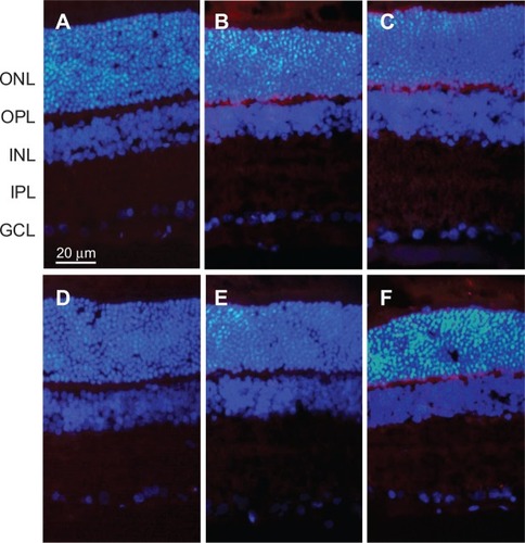
Figure 2 Immunohistochemistry staining of GFAP expressed in retinas at 2 weeks after intravitreal injection.
Notes: (A) Low group: intravitreal injection of 2.5 mg/0.1 mL PLGA/PLA microspheres; (B) medium group: intravitreal injection of 5 mg/0.1 mL PLGA/PLA microspheres; (C) high group: intravitreal injection of 10 mg/0.1 mL PLGA/PLA microspheres; (D) EPO group: intravitreal injection of 5 mg/0.1 mL EPO–dextran PLGA/PLA microspheres; (E) PBS group: intravitreal injection of 0.1 mL 0.01 M PBS; (F) normal group: normal retinas received no intravitreal injection. GFAP expression was mainly localized in the inner limiting membrane in all groups without any differences between groups.
Abbreviations: EPO, erythropoietin; GCL, ganglion cell layer; GFAP, glial fibrillary acidic protein; INL, inner nuclear layer; IPL, inner plexiform layer; ONL, outer nuclear layer; OPL, outer plexiform layer; PBS, phosphate-buffered saline; PLGA/PLA, poly(lactic-co-glycolic acid)/poly(lactic-acid).
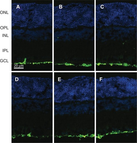
Figure 3 HE staining of structure changes in retinas at 2 weeks after intravitreal injection.
Notes: (A) Low group: intravitreal injection of 2.5 mg/0.1 mL PLGA/PLA microspheres; (B) medium group: intravitreal injection of 5 mg/0.1 mL PLGA/PLA microspheres; (C) high group: intravitreal injection of 10 mg/0.1 mL PLGA/PLA microspheres; (D) EPO group: intravitreal injection of 5 mg/0.1 mL EPO–dextran PLGA/PLA microspheres; (E) PBS group: intravitreal injection of 0.1 mL 0.01 M PBS; (F) normal group: normal retinas received no intravitreal injection. No obvious structure changes were detected in the retina in the injected groups compared with the intact eyes.
Abbreviations: EPO, erythropoietin; GCL, ganglion cell layer; HE, hematoxylin and eosin; INL, inner nuclear layer; IPL, inner plexiform layer; ONL, outer nuclear layer; OPL, outer plexiform layer; PBS, phosphate-buffered saline; PLGA/PLA, poly(lactic-co-glycolic acid)/poly(lactic-acid); PR, photoreceptor; RPE, retinal pigment epithelium.
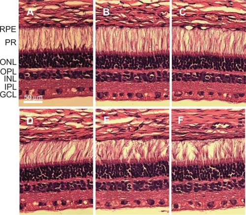
Figure 4 TEM scanning for ultrastructure changes of outer and inner segment of photoreceptors in retinas at 2 weeks after intravitreal injection.
Notes: (A) Low group: intravitreal injection of 2.5 mg/0.1 mL PLGA/PLA microspheres; (B) medium group: intravitreal injection of 5 mg/0.1 mL PLGA/PLA microspheres; (C) high group: intravitreal injection of 10 mg/0.1 mL PLGA/PLA microspheres; (D) EPO group: intravitreal injection of 5 mg/0.1 mL EPO–dextran PLGA/PLA microspheres; (E) PBS group: intravitreal injection of 0.1 mL 0.01 M PBS; (F) normal group: normal retinas received no intravitreal injection. No significant morphologic changes were detected in the outer segment disc and inner segment of mitochondria in the groups.
Abbreviations: EPO, erythropoietin; IS, photoreceptor inner segment; OS, photoreceptor outer segment; PBS, phosphate-buffered saline; PLGA/PLA, poly(lactic-co-glycolic acid)/poly(lactic-acid); TEM, transmission electron microscope.
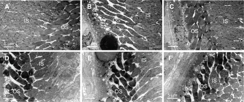
Figure 5 TEM scanning for ultrastructure changes of retinal ganglion cells at 2 weeks after intravitreal injection. Retinal ganglion cells are identified by arrows.
Notes: (A) Low group: intravitreal injection of 2.5 mg/0.1 mL PLGA/PLA microspheres; (B) medium group: intravitreal injection of 5 mg/0.1 mL PLGA/PLA microspheres; (C) high group: intravitreal injection of 10 mg/0.1 mL PLGA/PLA microspheres; (D) EPO group: intravitreal injection of 5 mg/0.1 mL EPO–dextran PLGA/PLA microspheres; (E) PBS group: intravitreal injection of 0.1 mL 0.01 M PBS; (F) normal group: normal retinas received no intravitreal injection. No distinct morphologic changes were detected in retinal ganglion cells in the injected eyes compared with normal retinas.
Abbreviations: EPO, erythropoietin; PBS, phosphate-buffered saline; PLGA/PLA, poly(lactic-co-glycolic acid)/poly(lactic-acid); TEM, transmission electron microscope.
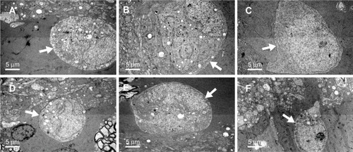
Figure 6 Scotopic ERG of rabbits at 2 weeks after intravitreal injection.
Notes: (A) Low group: intravitreal injection of 2.5 mg/0.1 mL PLGA/PLA microspheres; (B) medium group: intravitreal injection of 5 mg/0.1 mL PLGA/PLA microspheres; (C) high group: intravitreal injection of 10 mg/0.1 mL PLGA/PLA microspheres; (D) EPO group: intravitreal injection of 5 mg/0.1 mL EPO–dextran PLGA/PLA microspheres. No significant scotopic amplitudes were influenced by the injected microspheres compared with the intact eyes.
Abbreviations: a, A-wave of the ERG; b, B-wave of the ERG; EPO, erythropoietin; ERG, electroretinography; OD, the injected right eye; OS, the intact left eye; PLGA/PLA, poly(lactic-co-glycolic acid)/poly(lactic-acid).
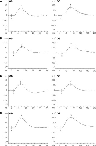
Figure 7 Photopic ERG of rabbits at 2 weeks after intravitreal injection.
Notes: (A) Low group: intravitreal injection of 2.5 mg/0.1 mL PLGA/PLA microspheres; (B) medium group: intravitreal injection of 5 mg/0.1 mL PLGA/PLA microspheres; (C) high group: intravitreal injection of 10 mg/0.1 mL PLGA/PLA microspheres; (D) EPO group: intravitreal injection of 5 mg/0.1 mL EPO–dextran PLGA/PLA microspheres. No significant photopic amplitudes were influenced by the injected microspheres compared with the intact eyes.
Abbreviations: a, A-wave of the ERG; b, B-wave of the ERG; EPO, erythropoietin; ERG, electroretinography; OD, the injected right eye; OS, the intact left eye; PLGA/PLA, poly(lactic-co-glycolic acid)/poly(lactic-acid).
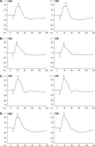
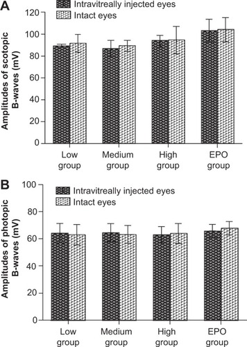
Figure 8 Scotopic (A) and photopic (B) ERG of rabbits at 2 weeks after intravitreal injection. Low group: intravitreal injection of 2.5 mg/0.1 mL PLGA/PLA microspheres; medium group: intravitreal injection of 5 mg/0.1 mL PLGA/PLA microspheres; high group: intravitreal injection of 10 mg/0.1 mL PLGA/PLA microspheres; EPO group: intravitreal injection of 5 mg/0.1 mL EPO–dextran PLGA/PLA microspheres. No statistically significant differences were observed between the intravitreally injected eyes and the intact eyes in all groups.
Abbreviations: EPO, erythropoietin; ERG, electroretinography; PLGA/PLA, poly (DL-lactic-co-glycolic acid)/poly(DL-lactic).