Figures & data
Figure 1 Schematic of the procedure for creating PLGA polymeric nanoparticles loaded with the PH-427 chemotherapeutic agent (PH-427-PNP). Emulsification in dichloromethane and water was facilitated by sonication, followed by solvent evaporation and purification using centrifugation.
Abbreviations: PLGA, poly(lactic-co-glycolic acid); PVA, poly(vinyl alcohol); PNP, PLGA polymeric nanoparticles.
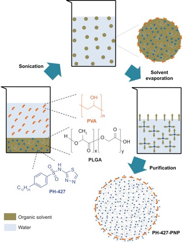
Figure 2 Characterization of the nanoparticles.
Notes: (A) Scanning electron microscopy images show a smooth surface for PLGA polymeric nanoparticles and drug-loaded PH-427-PNP. (B) Dynamic light scattering spectra of PNP and PH-427-PNP were used to determine the average diameter and polydispersity index of each nanoparticle, based on an average of ten measurements. (C) Experimental release of PH-427 from the PNP was performed in phosphate-buffered saline at pH 7.4 and 37°C, and then fit to a model that evaluates initial burst and slow relaxation of drug release.
Abbreviation: PLGA, poly(lactic-co-glycolic acid); PNP, poly(lactic-co-glycolic acid) polymeric nanoparticles.
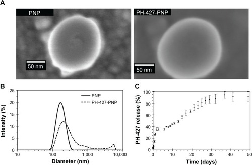
Figure 3 Fluorescence images show that Rhodamine-PNP were internalized in MiaPaCa-2 pancreatic cancer cells following 1 hour and 24 hours of treatment of the cells. Cells were visualized by detecting a 4′,6-diamidino-2-phenylindole (DAPI) stain (top), and Rhodamine-labeled PNP were visualized by detecting Rhodamine (bottom).
Abbreviation: PNP, poly(lactic-co-glycolic acid) polymeric nanoparticles.

Figure 4 Relative in vitro cell survival following drug treatments.
Notes: (A, B) BxPC3 and MiaPaCa-2 cell lines were treated once with PH-427 alone, and then evaluated with a methylthiazole tetrazolium (MTT) assay each day for 7 consecutive days. The results showed a lower therapeutic response for MiaPaCa-2 cells than for BxPC3 cells (P<0.001). (C, D) BxPC3 and MiaPaCa-2 cell lines were treated once with PNP loaded with the PH-427 chemotherapeutic agent (PH-427-PNP). The concentration of PH-427 was assumed to be the concentration recorded following the biphasic release effect. The percent cell survival was measured daily with a standard MTT microcytotoxicity assay for 7 consecutive days. These results showed that encapsulating PH-427 in the PNP improved the therapeutic efficacy in MiaPaCa-2 pancreatic cancer (B versus D; P<0.01), and had the same therapeutic efficacy against BxPC3 pancreatic cancer (A versus C; P>0.05), relative to treatment with PH-427 alone. (E, F) Results for C and D were re-evaluated by assuming that the concentration of PH-427 was the total concentration of drug in the PNP that was added to the cell system. Encapsulating PH-427 in the PNP did not improve therapeutic efficacy against MiaPaCa-2 pancreatic cancer (B versus F; P>0.05), and inclusion of PNP reduced the therapeutic efficacy against BxPC3 pancreatic cancer (A versus E; P<0.01). The error bars in each graph represent the standard deviation of each measurement (n=4).
Abbreviation: PNP, poly(lactic-co-glycolic acid) polymeric nanoparticles.
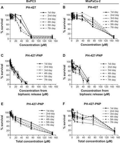
Figure 5 In vivo BLI following drug treatment.
Notes: (A) Bioluminescence images at 7 weeks after initiating treatment showed high average BLI signal amplitude for control mice and mice treated with PH-427, while low average BLI signal amplitude was observed in mice treated with PNP loaded with the PH-427 chemotherapeutic agent (PH-427-PNP). The average BLI signal and number of metastases to the liver determined from ex vivo assessments are shown below each image. (B) A quantitative comparison of average BLI signal amplitudes between the control group, the PH-427-treated group, and the PH-427-PNP-treated group. The differences between the PH-427-PNP-treated group and the other groups were statistically significant (P<0.001). (C) Ex vivo pancreatic tumor volumes were measured using calipers. The error bars in each graph represent the standard deviation of each measurement.
Abbreviations: BLI, bioluminescence imaging; Mets, metastases; PNP, poly(lactic-co-glycolic acid) polymeric nanoparticles.
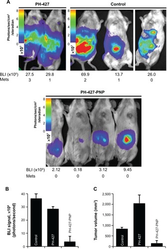
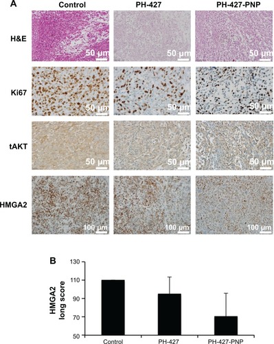
Figure 6 (A) In vivo inhibition of tumor proliferation and AKT. Representative staining is shown for H&E, Ki-67, tAKT, and high-mobility group AT-hook 2 (HMGA2) of control, PH-427-treated, and PH-427-PNP-treated mice (PH-427-PNP, PNP loaded with the PH-427 chemotherapeutic agent). (B) Long scores for HMGA2 staining for control, PH-427, and PH-427-PNP. The values are the means and the error bars are the standard errors (n=2). All images were acquired with 40× magnification.
Abbreviations: H&E, hematoxylin and eosin; PNP, poly(lactic-co-glycolic acid) polymeric nanoparticles; tAKT, total AKT.