Figures & data
Figure 1 Expression of SPINDOC in pan-cancer samples and its effect on prognosis. (A and B) SPINDOC expression was elevated in 18 types of malignant tumors compared with normal tissue. (C) Univariate analysis showed that SPINDOC could be a risk factor for prognosis in LIHC, KIRP and PCPG tumor. (D and E) High expression of SPINDOC can lead to poor prognosis in patients with KIRP and PCPG tumors. *P<0.05;**P<0.01;***P<0.001.
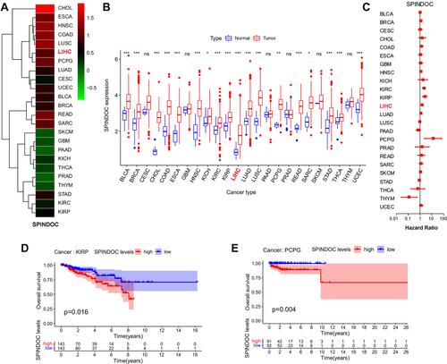
Figure 2 Genetic variation and methylation analysis of SPINDOC. (A) Types and frequency of genetic changes in SPINDOC. (B) Types, sites and proportions of SPINDOC mutations. (C and D) mRNA expression of SPINDOC is associated with mutations, and mutations lead to poor prognosis in patients. (E) Relationship between SPINDOC gene expression and methylation.
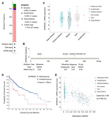
Figure 3 The gene expression level of SPINDOC in HCC and its potential effects on the clinical outcome of patients. (A) Patients with high SPINDOC expression displayed a poor prognosis; (B) SPINDOC was highly expressed in HCC patients younger than 60 years old; (C) The expression of SPINDOC increased with the clinical stage, (D) histological grade and (E) T stage of HCC. (F–I) GSEA analysis to identify the various cancer-related pathways where SPINDOC was enriched. (F) tumor-related pathways, (G) WNT signaling cascade, (H) adhesion junction, (I) cell cycle.
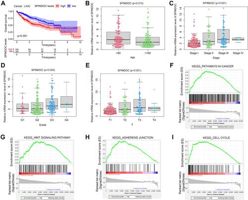
Table 1 Univariate and Multivariate Analysis of the Clinical Characteristics of HCC Patients and the Possible Correlation Between SPINDOC Gene Expression and Patient’s Survival Status
Figure 4 mRNA and protein expressions of SPINDOC in HCC cells. The proliferation and apoptosis ability of HCC cells after SPINDOC gene was knockdown. (A and B) mRNA and protein expressions of SPINDOC was markedly up-regulated in HCC cells; (C and F) Knockdown of SPINDOC gene can significantly attenuate colony forming ability of HCC cells; (D–E) Knockdown of SPINDOC can promote apoptosis of HCC cells. (G). Knockout of SPINDOC gene significantly reduced the survival rate of HCC cells at 48h and 72h respectively. **P<0.01.
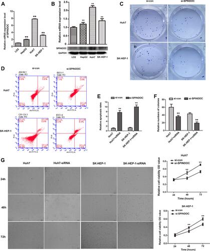
Figure 5 SPINDOC can promote the migration as well as invasion of HCC cells and can activate the Wnt/β-catenin signaling pathway. (A and B) Knocking down the expression of SPINDOC gene can inhibit the wound healing; (C–F) reduce the invasion and migration ability of HCC cells; (G) The expression of SPIN1, Wnt1, ß-catenin and cyclin D1 in HCC cells after SPINDOC gene knockdown decreased, but the expression of AXIN2 was found to be increased. **P<0.01.
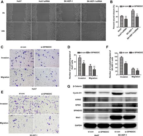
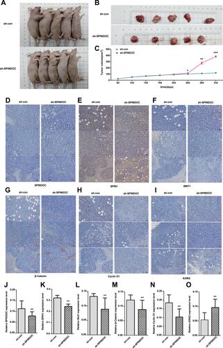
Figure 6 Knockdown of SPINDOC inhibited HCC growth and Wnt/β-catenin signaling pathway in nude mice. (A and B) Subcutaneous tumor growth was observed in both the control group and SPINDOC knockout group. (C) The growth of subcutaneous tumor in the nude mice after SPINDOC knockout was significantly smaller than that in the control group after 29 days (*P<0.05;**P<0.01;***P<0.001). (D–I) Immunohistochemical staining showed that protein expression levels of SPINDOC. (D and J) SPINDOC, (E and K) SPIN1, (F and L) Wnt1, (G and M) β-catenin, (H and N) cyclin D1 in SPINDOC-knockout group were lower than those in the control group, while the protein expression levels of AXIN2 (I and O) were higher than those in control group.**P<0.01.