Figures & data
Figure 1 (A) The structure of scdsFv version of HA22LR. The 2-chain disulfide- and peptide-(linker 1) linked Fv of an antibody targeting CD22 is combined with the domain III of native PE by the linker which bears Furin-cleavage site (linker 2) to create an immunotoxin. The Fv portion sequence is shown. Residue numbering is based on the Kabat numbering scheme.Citation22 The CDR regions are defined according to Kabat et al. (underlined) and IMGT23 (boldface). Alanine-scanned CDR residues are shown in red. (B) Activities of dsFv-form (circles and dotted line) and scdsFv-form (squares and solid line) of HA22-LR on CD22-positive cells. The cytotoxicity was measured by WST-8 in triplicate 3 times. Typical cytotoxic curves are shown. Data are expressed as the mean ± SD. IT, immunotoxin.
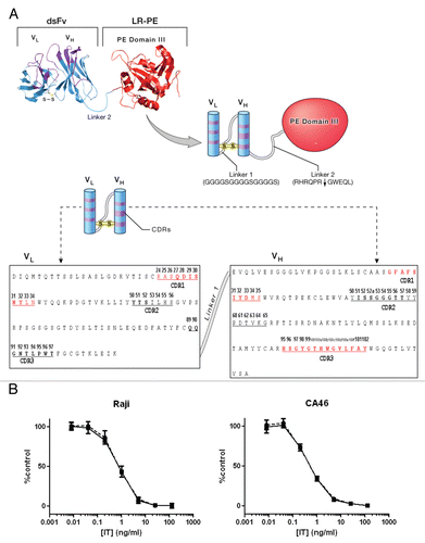
Figure 2 SDS-PAGE analysis of purified immunotoxins. Ten µg of purified immunotoxins were loaded per lane. Gel picture of 10 immunotoxins is shown as representative of the size and purity of all immunotoxins used in this study.
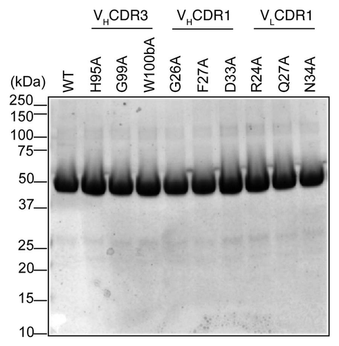
Figure 3 Ribbon model of position VL34 of HA22-Fv. VL34 is buried and located at the interface of VL and VH.
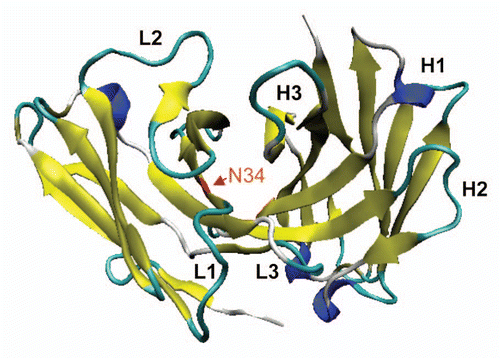
Figure 4 Characterization of N34A mutant. (A) Affinities of WT scdsFv-HA22-LR (circles and dotted line) and its N34A mutant (squares and solid line) to CD22-positive Daudi cells. Affinities were measured by FACS. Briefly, pre-fixed Daudi cells were incubated with immunotoxins at 4°C overnight. Bound immunotoxins were detected with anti-LR-PE mouse polyclonal antibodies and PE-labeled goat anti-mouse IgG. Anti-mesothelin immunotoxin SS1PCitation6 (triangles and dotted line) was used as a negative control. Mean fluorescence intensities are shown. Each assay was performed in triplicate. Data are expressed as the mean ± SD. (B) Specific cytotoxic activities of WT scdsFv-HA22-LR (circles and dotted line) and its N34A mutant (squares and solid line) on CD22-positive cells. The cytotoxicity was measured by WST-8 in triplicate at least nine times. Typical cytotoxic curves are shown. Data are expressed as the mean ± SD. We analyzed a total of 8 cell lines, and their IC50 concentrations are shown in .
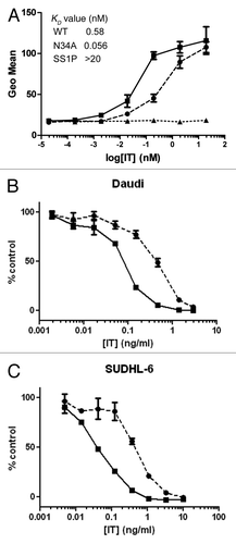
Table 1 Specific cytotoxic activities of mutants in CDRs
Table 2 Specific cytotoxic activities of WT and N34A mutant of scdsFvHA22LR on various cell lines