
Abstract
Background
Cigarette smoke (CS) increases oxidative stress (OS) in the lungs. Pomegranate juice (PJ) possesses potent antioxidant activities, attributed to its polyphenols. This study investigates the effects of PJ on the damaging effects of CS in an animal model and on cultured human alveolar cells (A549).
Methods
Male C57BL/6J mice were divided into the following groups: Control, CS, CS + PJ, and PJ. Acute CS exposure was for 3 days, while chronic exposure was for 1 and 3 months (5 days of exposure/week). PJ groups received daily 80 μmol/kg via bottle, while other groups received distilled water. At the end of the experiments, different parameters were studied: 1) expression levels of inflammatory markers, 2) apoptosis, 3) OS, and 4) histopathological changes. In vitro, A549 cells were pretreated for 48 hours with either PJ (0.5 μM) or vehicle. Cells were then exposed to increasing concentrations of CS extracted from collected filters. Cell viability was assessed by counting of live and dead cells with trypan blue staining.
Results
Acutely, a significant increase in interleukin (IL)-1β, IL-6, and tumor necrosis factor (TNF)-α expression, apoptosis, and OS was noted in CS when compared to Control. PJ significantly attenuated the expression of inflammatory mediators, apoptosis, and OS. Chronically (at 1 and 3 months), increased expression of TNF-α was observed, and lung sections demonstrated emphysematous changes when compared to Control. PJ supplementation to CS animals attenuated the increased expression of TNF-α and normalized lung cytoarchitecture. At the cellular level, CS extract reduced cellular proliferation and triggered cellular death. Pretreatment with PJ attenuated the damaging effects of CS extract on cultured human alveolar cells.
Conclusion
The expression of inflammatory mediators associated with CS exposure and the emphysematous changes noted with chronic CS exposure were reduced with PJ supplementation. In vitro, PJ attenuated the damaging effects of CS extract on cultured human alveolar cells.
Introduction
Cigarette smoke (CS) is a major risk factor for chronic bronchitis, asthma, and lung cancer. It also remains the main culprit in the pathogenesis of chronic obstructive pulmonary disease (COPD).Citation1–Citation3 CS exposure is known for tipping the oxidative balance in the respiratory system, culminating in an oxidative stress (OS) status.Citation4 CS contains >5,000 different chemicals, many of which are oxidants. It is estimated that each puff may contain >1015 free radicals that deplete endogenous antioxidants and tilt the delicate balance in favor of an OS.Citation4–Citation6 Increased OS is well described in patients with COPD;Citation7 increased levels of H2O2 and 8-isoprostane were demonstrated in the exhaled breaths of COPD patients, and increased markers of OS were also present in lung cells of COPD patients.Citation8–Citation10 An increase in the levels of nitrotyrosine, an indicator of cellular damage secondary to free radicals, has also been noted in sputum leukocytes and lung tissue of COPD patients when compared to healthy subjects.Citation11,Citation12
Punica granatum L. (Punicaceae) is usually consumed as pomegranate juice (PJ). Polyphenols, present in PJ, possess potent antioxidants, which may contribute to its antiatherosclerotic and anti-inflammatory properties.Citation13–Citation16 Dietary supplementation of polyphenols may potentially play a therapeutic role in protecting against CS-induced OS.Citation17 Polyphenols act as scavengers of oxygen radical and hydroxyl radical molecules and increase the levels of the antioxidant glutathione by the induction of glutamate cysteine ligase.Citation4 The combination of polyphenols with other phytochemicals such as ellagic acid synergistically enhances the superior antioxidant properties of PJ.Citation18 In addition, there is the added benefit of the anti-inflammatory properties of polyphenols due to the inhibition of nuclear factor kappa B (NF-κB) expression/activation, interleukin (IL)-8 release, cyclooxygenase-2, and heme oxygenase-1.Citation19,Citation20
This study examined whether supplementation of PJ attenuates the damaging effects of acute and chronic CS exposure both in the lungs of an animal model and at the cellular level.
Methods
In vivo study
The Institutional Animal Care and Use Committee of the American University of Beirut approved this study. Four-month-old adult male C57BL/6J mice (22–25 g body weight) were subjected to a 12-hour dark/light cycle. Temperatures of the room and chambers were maintained at 22°C–24°C, and animals were allowed unlimited access to water and standard rodent chow except when animals were placed in the exposure apparatus. The CS exposure apparatus (ONARES; CH Technologies, Westwood, NJ, USA) consisted of a smoke generator, mixing/conditioning chamber, and a 12-port “nose–only” rodent exposure carousel. One port of the carousel was dedicated for sampling analysis and the remaining eleven ports were used for animal exposure. Animals were divided into four groups: Control, CS, CS + PJ, and PJ. Each group consisted of eleven animals, and all animals were acclimated to retainers for 1 week before initiating exposure to laboratory air or CS. Mice were then positioned in retainers and placed into the holes of the carousel. Animals received a continuous flow of CS or room air into the airways via the “nose–only” delivery system. CS was generated from 3R4F cigarettes (University of Kentucky, Lexington, KY, USA) with 0.9 mg total particulate matter (TPM), 9.4 mg tar, and 0.726 mg nicotine per cigarette. As described previously, the machine was set at one puff every minute, with duration of 2 seconds per puff and a volume of 35 mL per puff.Citation21
The sampling system consisted of a vacuum pump attached to one port of the carousel, which drew the diluted aerosol at 1 L/min (controlled by a critical orifice) through a 47 mm fiberglass filter disk (CH Technologies). Filters were replaced every 30 minutes. The TPM per cubic meter (ΔW) was determined gravimetrically by weighing each filter before CS exposure using an analytical balance. TPM concentration was calculated by dividing ΔW over the time and air sampling rate (1 L/min).
The study was performed at three different time points. The acute exposure time setup was deduced from the literature and was set for 3 consecutive days.Citation22 Animals were exposed to CS or laboratory air twice daily (9 am and 2 pm) for 3 hours each. The calculated TPM for the acute exposure was approximately 100 TPM/m3. The chronic exposure setup consisted of two time points (1 and 3 months). Animals were exposed for two sessions of CS (9 am and 2 pm) per day for 5 days/week. Each session, however, lasted for 1 hour and the calculated TPM was approximately 100 TPM/m3.
At the conclusion of the experiment, animals were anesthetized and exsanguinated by severing the aorta. The diaphragm was dissected to allow free lung expansion. The lower lobe of left lung was excised for pulmonary water content evaluation. The left upper lobe was fixed in formalin for terminal deoxynucleotidyl transferase-mediated dUTP nick-end labeling (TUNEL) assay, OS measurements, and pathology examination. The remaining right lung lobes were frozen for RNA extraction.
Pomegranate juice (PJ) processing and administration
PJ concentrate (Wonderful variety; POM Wonderful, Los Angeles, CA, USA), utilized in this study, was administered via bottle to the CS + PJ and PJ groups. PJ was initiated 1 week before CS or room air exposure and was maintained throughout the experiment. Animals received 80 μmol/kg/day of PJ, while the Control and CS groups received free water. The dose of PJ supplementation was deduced from previous studies.Citation23
Wet-to-dry lung weight
The left lower lobe was weighed and then placed in a 95°C oven to dry for 2 days. The dry tissue was weighed, and the wet-to-dry (W/D) ratio was then calculated.
Transcription expression of IL-1, IL-6, and tumor necrosis factor-α
Changes in the inflammatory mediators’ transcriptional levels were assessed using the reverse transcriptase–polymerase chain reaction (PCR) method. RNA was extracted using the TRIzol method (Invitrogen, Carlsbad, CA, USA) as described before.Citation24 Briefly, 1 mL of TRIzol reagent was used per 50–100 mg of tissue sample, followed by chloroform extraction. RNA samples were precipitated and stored at −80°C. RNA was quantified using the 260/280 nm absorbance ratio method. Total RNA (5 μg) was reverse-transcribed into first–strand complementary DNA (cDNA). Real-time-PCR was performed using the iCycler (Bio-Rad Laboratories, Hercules, CA, USA) with SYBR Green. Specific primers (Tib-Molbiol, Berlin, Germany) were used to assess the expression of the inflammatory mediators in these tissues (IL-1β: Forward CACCTCTCAAGCAGAGCA-CAG, Reverse GGGTTCCATGGTGAAGTCAAC; IL-6: Forward TCCTACCCCAACTTCCAATGCTC, Reverse TTGGATGGTCTTGGTCCTTAGCC; tumor necrosis factor-α [TNF-α]: Forward AATGGGCTCCCTCTCAT-CAGTTC, Reverse TCTGCTTGGTGGTTTGCTACGAC). PCR products and their corresponding melting temperatures were analyzed using the iQ5 Optical System Software (Bio-Rad Laboratories). Correction for loading was achieved by subtracting for local background and normalizing against the cDNA levels of the glyceraldehyde-3-phosphate dehydrogenase (GAPDH) housekeeping gene (GAPDH: Forward GTATTGGGCGCCTGGTCACC, Reverse CGCTCCTGGAAGATGGTGATGG).
Assessment of oxidative stress
Dihydroethidium (DHE) (Molecular Probes; Thermo Fisher Scientific, Waltham, MA, USA) (10 μmol/L dissolved in dimethyl sulfoxide) was applied to lung sections and incubated in a light-protected humidified chamber at 37°C for 15 minutes. Fluorescent images of ethidium-stained tissue were scanned for signal with a scanning confocal microscope (Zeiss, Oberkochen, Germany). Ethidium bromide was excited at 488 nm and emission fluorescence was detected at 560 nm.
Assessment of apoptosis
TUNEL assay was used to monitor the extent of DNA fragmentation.Citation23 Fluorescein-conjugated dUTP incorporated in nucleotide polymers was detected and analyzed using fluorescence microscopy (LSM 410; Zeiss). Positive and negative controls were used to verify the specificity of the TUNEL assay. TUNEL-positive nuclei were distinguished from the TUNEL-negative nuclei by counterstaining with Hoechst 33258.
Lung histology
The upper lobe of the left lung was fixed in 10% buffered formalin, embedded in paraffin, serially sectioned, and stained with hematoxylin and eosin (H&E). A board-certified pathologist, blinded to the different animal groups, evaluated the histopathologic findings under light microscopy (Axio Observer; Zeiss) and determined the degree of lung injury based on degree of inflammatory cell infiltration, alveolar edema, and emphysema.
Quantification of emphysema
Pulmonary mean linear intercept (Lm), an indicator of air space size, was calculated for each sample based on random fields observed at a total magnification of ×200 using a crossline.Citation24 The total length of the crossline divided by the number of the alveolar walls intersecting the test lines was defined as Lm.
In vitro study
Preparation of CS extract
CS extract (CSE) was obtained from filters collected during the animal studies. On the basis of the weight of the TPM collected by the filter, Dulbecco’s Modified Eagle’s Medium (DMEM) incomplete medium was added to yield a final concentration of 10 mg/mL. All recovered media were then mixed together and sterilized using 0.22 μm filters (Costar; Corning, NY, USA).
Cell culture
A549 cells were grown in DMEM high glucose (4.5 g/L) culture media and supplemented with penicillin-G 100 U/mL, streptomycin 100 μg/mL (Gibco-BRL, Paisley, UK), and 10% fetal bovine serum (Sigma-Aldrich Co, St Louis, MO, USA). Cells were seeded in 24-well plates at a density of 60,000 cells per well. Cells receiving PJ supplementation were incubated with 0.5 μM of PJ for 48 hours before exposure to CSE or placebo sterile water. Exposure to CSE for 24 hours was initiated by mixing CSE (which was prepared as stock solutions at concentration of 10 mg/mL) and complete media to the desired final concentration (0.5, 1, 2, 4, and 8 mg/mL). Images were then taken using a light microscope (Axio Observer; Zeiss). Cells were then counted using trypan blue dye to differentiate between dead and live cells. The Institutional Review Board of the American University of Beirut did not require approval be sought for the use of the cell lines in this study, as they are available commercially.
Measurement of ROS
A549 cells were seeded in 24-well plates containing cover slips at a density of 60,000 cells per well. Cells receiving PJ supplementation were incubated with 0.5 μM of PJ for 48 hours before exposure to CSE or placebo sterile water. CSE stock solutions were prepared at a concentration of 10 mg/mL and exposure to CSE for 24 hours was initiated by mixing CSE and complete media to the desired final concentration (0.5, 1, 2, 4, and 8 mg/mL). Cells were then washed with 1× phosphate-buffered saline, pH =7.4. Cells were incubated with 300 μL of DHE (10 μmol/L dissolved in dimethyl sulfoxide and incubated in light-protected humidified chamber at 37°C) for 15 minutes. DHE was then removed and 4% formaldehyde was added for 30 minutes. The cover slips were then mounted on glass slides using ProLong Antifade. Images were taken using a laser scanning confocal microscope (LSM-710; Zeiss).
Results
Acute CS exposure
There was no significant change in the weight of animals between the different groups throughout the different experiments (acute and chronic). There was no difference in W/D ratio, the expression of inflammatory mediators, apoptotic activity, or OS between Control and PJ. A statistically significant increase in mean W/D in CS was noted when compared to Control (P=1.1×10⁻⁷). PJ supplementation attenuated the increase in W/D observed in CS (P=1×10⁻⁶) (). The expression of IL-6, IL-1β, and TNF-α was significantly increased in CS when compared to Control (P=0.04, 0.01, and 0.004, respectively) and PJ supplementation to CS animals significantly reduced the expression of inflammatory mediators noted in CS (). The lungs of CS animals demonstrated a significant increase in OS and in the number of TUNEL-positive apoptotic nuclei when compared to Control (). Again, PJ supplementation significantly attenuated the increased OS and apoptotic activity noted in CS.
Figure 1 Wet-to-dry ratio (W/D) and expression of inflammatory mediators after acute exposure to CS.
Notes: Mean W/D (A), transcriptional expression of IL-1β (B), IL-6 (C), and TNF-α (D) of Control, acute CS exposure (CS), CS + PJ, and PJ. Error bars represent SE. Asterisks indicate statistically significant associations (P<0.05).
Abbreviations: CS, cigarette smoke; IL, interleukin; PJ, pomegranate juice; SE, standard error; TNF, tumor necrosis factor.
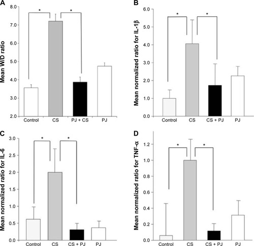
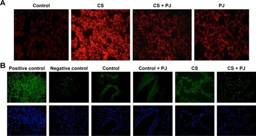
Figure 2 PJ attenuates CS-induced ROS formation and apoptosis.
Abbreviations: CS, cigarette smoke; DHE, dihydroethidium; PJ, pomegranate juice; ROS, reactive oxygen species; TUNEL, terminal deoxynucleotidyl transferase-mediated dUTP nick-end labeling.
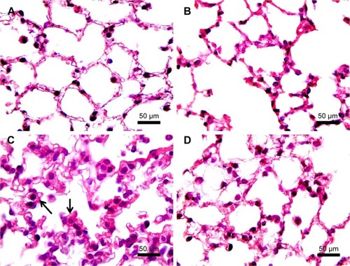
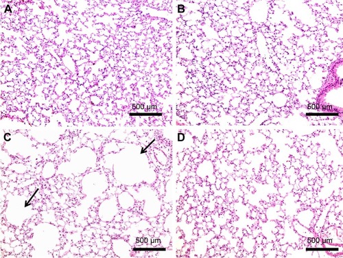
H&E staining of lung sections of CS animals revealed the increased presence of inflammatory cells (macrophages and lymphocytes) across the bronchioles and in lung paren-chyma. Edematous and thickened alveolar walls were also observed. CS animals treated with PJ displayed normal alveolar structure with minimal infiltration of inflammatory cells and swelling of the alveoli ().
Figure 3 H&E examination under light microscopy (original magnification: ×100).
Abbreviations: CS, cigarette smoke; H&E, hematoxylin and eosin; PJ, pomegranate juice.
One-month CS exposure
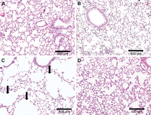
After 1 month of CS exposure, only TNF-α demonstrated persistent increased expression in CS when compared to Control (P=0.04) (). PJ supplementation in the CS + PJ group normalized the increased expression of TNF-α observed in CS. Histologically, subtle changes suggestive of early enlargement of the airspaces, accompanied with limited destruction of the normal alveolar architecture (increases of airspace enlargement), were noted in CS. In contrast, CS treated with PJ displayed normal alveolar structure ().
Figure 4 Mean transcriptional expression of TNF-α after chronic exposure.
Notes: Significantly increased levels of TNF-α were observed in CS animals at 1 month (A) and 3 months (B) of CS. The increased levels of TNF-α were significantly attenuated with PJ supplementation. Error bars represent SE. Asterisks indicate statistically significant associations (P<0.05).
Abbreviations: CS, cigarette smoke; PJ, pomegranate juice; SE, standard error; TNF, tumor necrosis factor.
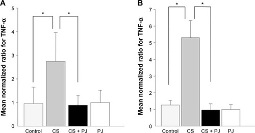
Figure 5 H&E examination under light microscopy at 1 month (original magnifications: ×20).
Abbreviations: CS, cigarette smoke; H&E, hematoxylin and eosin; PJ, pomegranate juice.
Three-month CS exposure
Similar to the results of 1-month CS exposure, increased expression of TNF-α was also observed in CS, which was attenuated with PJ supplementation (P=0.001) (). Histological evaluation after 3 months of CS, however, revealed significant emphysematous changes with enlargement of the airspaces, accompanied by the destruction of the normal alveolar architecture in CS (). Linear intercept data confirmed the significant increase in airspace size in CS lungs compared to Control lungs (P<0.0001). Chronic supplementation with PJ reversed the emphysema-tous changes noted histologically and attenuated the increase in Lm distance observed in the CS group (P<0.0001).
Figure 6 H&E examination under light microscopy at 3 months (original magnification: ×20).
Abbreviations: CS, cigarette smoke; H&E, hematoxylin and eosin; PJ, pomegranate juice.
In vitro study
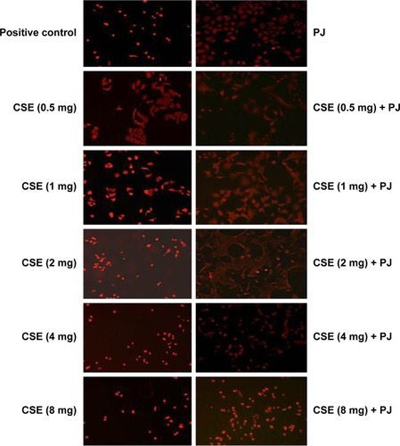
CSE reduced cellular proliferation, when compared to Control, in a dose-dependent manner. At 2 mg/mL of CSE exposure, cellular proliferation was completely arrested and cellular death was observed. Human alveolar cells, pretreated with PJ at a dose of 0.5 μM, demonstrated significant resistance to the effects of CSE and shifted cellular inhibition and death to higher doses of CSE. Arrest of cellular growth and cellular death were seen in CSE-only exposure at 2 mg/mL of CSE, whereas cellular proliferation was nearly unaffected and cells appeared healthy in PJ + CSE at the same concentration (). Similarly, a significant increase in OS in A549 cells was observed with increased concentrations of CSE. Again, PJ supplementation suppressed CS-induced reactive oxygen species (ROS) activity in A549 cells ().
Figure 7 Cellular morphology examination under light microscopy (original magnification: ×20).
Notes: Changes in cellular morphology were observed with increasing concentrations of CS extract. Cell shrinkage, rounding, and vacuoles were observed with CSE of concentrations ≥1 mg/mL.
Abbreviations: CS, cigarette smoke; CSE, CS extract; PJ, pomegranate juice.
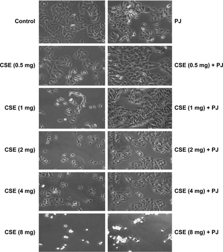
Figure 8 CSE-induced oxidative stress in A549 cells.
Abbreviations: CS, cigarette smoke; CSE, CS extract; DHE, dihydroethidium; PJ, pomegranate juice; ROS, reactive oxygen species.
Discussion
This study examined the damaging effects of CS in an animal model at different time points (acute and chronic) and in human alveolar cells. The “nose–only” delivery system, utilized in the animal study, delivered CS directly into the airways in a continuous, precise, and timely manner. This eliminated inconsistent CS exposure associated with whole-body exposure. As such, accelerated lung injury and early emphysematous changes were observed after 1 month of CS exposure.Citation25 Acute CS exposure was associated with a significant increase in W/D ratio, OS, cellular death, and a surge in inflammatory mediators, in association with infiltration by inflammatory cells.Citation26–Citation28 After 1 or 3 months of CS exposure, only the expression of TNF-α remained persistently elevated, and emphysematous changes with loss of alveolar sacs were noted at 1 month but were clearly evident at 3 months. The findings of persistent increased expression of TNF-α are consistent with the detrimental role of TNF-α in CS. TNF-α is known to prime neutrophils, on exposure to CS, resulting in an increase in its oxidative burst expression, leading to augmented lung damage. In mouse animal models similar to our model, TNF-α was noted to be central in CS-induced loss of alveoli and emphysema.Citation28–Citation30 At the cellular level, CSE, in a dose-dependent manner, inhibited cellular growth and induced cellular death of human alveolar cell cultures.
The study then examined the role of antioxidants in attenuating lung injury secondary to CS. This role is questioned due to recent conflicting, and rather disap po inting, animal and human studies that described limited – and possibly adverse – effects associated with exogenous antioxidant supplementation.Citation31–Citation33 Excessive administration of antioxidants suppresses total endogenous ROS formation and diminishes the capability of the recipient to kill bacteria and eliminate damaged or precancerous cells.Citation34 The results of this study supported the role of pomegranate as a powerful antioxidant in protecting the lungs from CS exposure. In vivo, PJ reversed and attenuated all the damaging effects of CS. Acutely, PJ supplementation attenuated the increase in W/D ratio, OS, apoptosis, and the surge of all inflammatory mediators associated with CS. Chronically, PJ attenuated the elevated expression of TNF-α and the emphysematous changes observed histologically with the increase in airspaces and the loss of alveoli. Finally, at the cellular level, PJ created significant resistance to the effects of CSE and higher doses of CSE concentration were needed to elicit similar results as in CSE-only cell cultures.
The success in achieving therapeutic outcomes with antioxidant supplementation is dependent on the choice, the dose of the antioxidant, and the timing of administration.Citation34,Citation35 PJ, utilized in this study, is an important source of powerful antioxidants that include anthocyanins, pelargonidin, and polyphenols. PJ possesses the added value of anti-inflammatory properties as well.Citation19,Citation20 As for the precise dose of daily PJ supplementation, based on previous studies, the daily dose of PJ was set at 80 μmol/kg/day.Citation23 Finally, the timing of administration is vital. In this study, PJ supplementation was initiated 1 week before the exposure to CS, priming animals with supplementary antioxidants needed to neutralize ROS generated from CS.
Emphysema is the hallmark of CS-induced lung injury.Citation3 This study demonstrated the favorable effects of antioxidant supplementation, represented by PJ, in limiting the damaging effects of CS and preventing the formation of emphysematous changes in the lung in an animal model. Further animal and human studies are needed to explore the beneficial role of antioxidants, with clear emphasis on the design of the study, and these need to precisely determine the dosing and timing of antioxidant administration in order to elicit a positive protective effect.
Author contributions
All authors contributed toward data analysis, drafting and critically revising the paper, gave final approval of the version to be published, and agree to be accountable for all aspects of the work.
Acknowledgments
This study was supported by the Mikati Foundation, Beirut, Lebanon, and the Medical Practice Plan and the University Research Board at the American University of Beirut, Beirut, Lebanon.
Disclosure
The authors report no conflicts of interest in this work.
References
- TamimiASerdarevicDHananiaNAThe effects of cigarette smoke on airway inflammation in asthma and COPD: therapeutic implicationsRespir Med2012106331932822196881
- U.S. Department of Health and Human ServicesThe Health Consequences of Smoking – 50 Years of Progress: A Report of the Surgeon GeneralAtlanta, GAU.S. Department of Health and Human Services, Centers for Disease Control and Prevention, National Center for Chronic Disease Prevention and Health Promotion, Office on Smoking and Health2015
- U.S. Department of Health and Human ServicesHow Tobacco Smoke Causes Disease: What It Means to YouAtlanta, GAU.S. Department of Health and Human Services, Centers for Disease Control and Prevention, National Center for Chronic Disease Prevention and Health Promotion, Office on Smoking and Health2015
- PasupathiPSaravananGFarookJOxidative stress bio markers and antioxidant status in cigarette smokers compared to nonsmokersJ Pharm Sci Res2009125562
- ForniLGMora-ArellanoVOPackerJENitrogen dioxide and related free radicals: electron-transfer reactions with organic compounds in solution containing nitrite or nitrateJ Chem Soc Perkin Trans1986216
- RahmanIAdcockIMOxidative stress and redox regulation of lung inflammation in COPDEur Respir J200628121924216816350
- BowlerRPBarnesPJCrapoJDThe role of oxidative stress in chronic obstructive pulmonary diseaseCOPD20041225527717136992
- AntczakACiebiadaMPietrasTExhaled eicosanoids and biomarkers of oxidative stress in exacerbation of chronic obstructive pulmonary diseaseArch Med Sci20128227728522662001
- FoschinoMPCarpagnanoGESpanevelloAInflammation, oxidative stress and systemic effects in mild chronic obstructive pulmonary diseaseInt J Immunopathol Pharmacol200720475376318179748
- KostikasKPapatheodorouGPsathakisKOxidative stress in expired breath condensate of patients with COPDChest200312441373138014555568
- LisetteIZKunzJBSimonaEBSmoking status and anti-inflammatory macrophages in bronchoalveolar lavage and induced sputum in COPDRespir Res2011123421426578
- IchinoseMSugiuraHYamagataSIncrease in reactive nitrogen species production in chronic obstructive pulmonary disease airwaysAm J Respir Crit Care Med20001622 pt 170170610934109
- LangleyPWhy a pomegranate?BMJ200032176291153115411061746
- El-NemrSEIsmailIARagabMChemical composition of juice and seeds of pomegranate fruitNahrung19907601606
- GilMITomas-BarberanFAHess-PierceBAntioxidant activity of pomegranate juice and its relationship with phenolic composition and processingJ Agric Food Chem200048104581458911052704
- FilomenaNSharonWLilachOLBeneficial effects of pomegranate juice on oxidation-sensitive genes and endothelial nitric oxide synthase activity at sites of perturbed shear stressProc Natl Acad Sci U S A20011021348964901
- ArtsICHollmanPCPolyphenols and disease risk in epidemiologic studiesAm J Clin Nutr2005811 suppl317S325S15640497
- SreekumarSSithulHMuraleedharanPPomegranate fruit as a rich source of biologically active compoundsBiomed Res Int2014201468692124818149
- BiswasSKMcClureDJimenezLACurcumin induces glutathione biosynthesis and inhibits NF-κB activation and interleukin-8 release in alveolar epithelial cells: mechanism of free radical scavenging activityAntioxid Redox Signal200571–2324115650394
- YunNKangJWLeeSMProtective effects of chlorogenic acid against ischemia/reperfusion injury in rat liver: molecular evidence of its antioxidant and anti-inflammatory propertiesJ Nutr Biochem201223101249125522209001
- CogginsCRA review of chronic inhalation studies with mainstream cigarette smoke, in hamsters, dogs, and nonhuman primatesToxicol Pathol200129555055711695572
- BondJAChenBTGriffithWCInhaled cigarette smoke induces the formation of DNA adducts in lungs of ratsToxicol Appl Pharmacol19899911611722727995
- HusariAKhayatABitarHAntioxidant activity of pomegranate juice reduces acute lung injury secondary to hyperoxia in an animal modelBMC Res Notes201421766425241213
- HusariAKhayatAAwdehHActivated protein C attenuates acute lung injury and apoptosis in a hyperoxic animal modelShock201033546747219851127
- BeckettELStevensRLJarnickiAGA new short-term mouse model of chronic obstructive pulmonary disease identifies a role for mast cell tryptase in pathogenesisJ Allergy Clin Immunol2013131375276223380220
- TakuboYGuerassimovAGhezzoHAlpha1-antitrypsin determines the pattern of emphysema and function in tobacco smoke-exposed mice: parallels with human diseaseAm J Respir Crit Care Med200216612 pt 11596160312471075
- KamiideYFuruyaMInomataNChronic exposure to cigarette smoke causes extrapulmonary abnormalities in ratsEnviron Toxicol Pharmacol201539286487025770835
- ChurgAWangRDTaiHTumor necrosis factor-alpha drives 70% of cigarette smoke-induced emphysema in the mouseAm J Respir Crit Care Med2004170549249815184206
- FriedrichsBNeumannUSchüllerJCigarette-smoke-induced priming of neutrophils from smokers and non-smokers for increased oxidative burst response is mediated by TNF-αToxicol In Vitro20142871249125824997298
- LiYTHeBWangYZExposure to cigarette smoke upregulates AP-1 activity and induces TNF-alpha overexpression in mouse lungsInhal Toxicol200921764164719235541
- BjelakovicGNikolovaDGluudLLAntioxidant supplements for prevention of mortality in healthy participants and patients with various diseasesCochrane Database Syst Rev2012143
- AlbanesDBeta-carotene and lung cancer: a case studyAm J Clin Nutr199969613451350
- WeissigVGuzman-VillanuevaDNanocarrier-based antioxidant therapy: promise or delusion?Expert Opin Drug Deliv201512111783179026119920
- JainMChandelNSRethinking antioxidants in the intensive care unitAm J Respir Crit Care Med2013188111283128524117139
- PoljsakBŠuputDMilisavIAchieving the balance between ROS and antioxidants: when to use the synthetic antioxidantsOxid Med Cell Longev2013201395679223738047